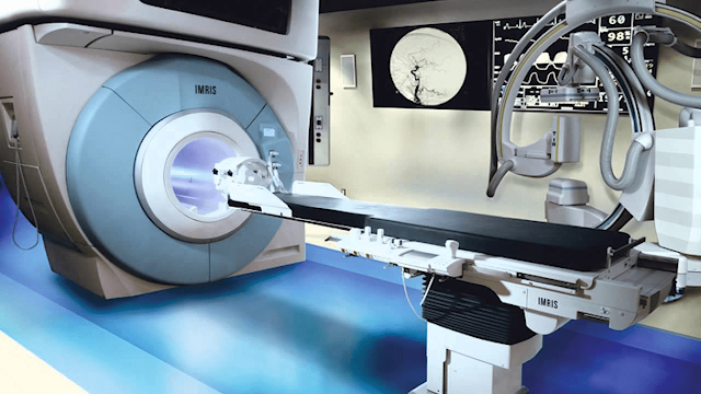
Intraoperative Imaging Useful To Provide Real-Time Visualization During Surgical Procedures
 |
| Intraoperative Imaging |
Intraoperative Imaging refers to the use of various imaging techniques during surgical procedures to provide real-time visualization and guidance to the surgeon. These techniques allow the surgeon to assess the extent of the disease, identify any abnormalities, and navigate complex anatomical structures, thus improving surgical outcomes.
The most common Intraoperative Imaging modalities are X-ray, ultrasound, computed
tomography (CT), magnetic resonance imaging (MRI), and fluoroscopy. Each of
these modalities has its advantages and limitations, and the choice of modality
depends on the type of surgery and the specific requirements of the surgeon.
Global
Intraoperative Imaging Market Is Estimated To Be Valued At US$ 2,868.7 Million In 2022 And
Is Expected To Exhibit A CAGR Of 6.4% During The Forecast Period (2022-2030).
X-ray is one of the oldest and most widely used
intraoperative imaging modalities. It uses electromagnetic radiation to create
images of the internal structures of the body. In surgery, X-ray is commonly
used to visualize bones, such as in orthopedic procedures. X-ray is fast,
non-invasive, and relatively inexpensive, but it has limited soft tissue
contrast and exposes the patient and surgical team to ionizing radiation.
Ultrasound is another commonly used Intraoperative Imaging modality that
uses high-frequency sound waves to create images of internal structures. It is
especially useful in surgeries involving the liver, pancreas, and other
abdominal organs. Ultrasound is fast, non-invasive, and does not expose the patient
to ionizing radiation, making it an attractive option for intraoperative
imaging.
CT and MRI are more advanced imaging modalities
that offer higher resolution and greater soft tissue contrast than X-ray and
ultrasound. CT uses X-rays and computer algorithms to generate detailed,
three-dimensional images of the body, while MRI uses a strong magnetic field
and radio waves to create images. Both CT and MRI provide highly detailed
images of the internal structures of the body, making them valuable tools in
complex surgical procedures. However, they are relatively slow and expensive,
and their use in the operating room requires specialized equipment and
expertise.


Comments
Post a Comment